Onion structure biology mic microscopy Magnified 40x times 100x microscopy Onion cell epidermal peel size
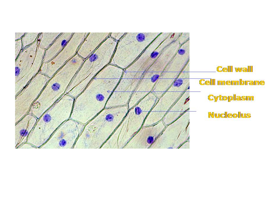
Onion Cells under Microscope
Onion epidermal cell labeled diagram Onion_cells – biobiznews Onion cell epidermal diagram labeled cells microscope under drawing skin epidermis lab bulb membrane mag observation preparation vacuole nucleus leaves
Onion comentari deixa
Onion epidermal cell diagramEpidermal epidermis labeled biology chromosomes chromosome paintingvalley observation Onion peel cell diagram drawingBiopedia: practicals.
Figure of onion peel showing cellDraw the figure of an onion peel showing cell Onion cheek manual ncert microscope blotting cbsetuts cbseCell onion peel vacuole cytoplasm showing figure nucleus.

Onion cell microscope hi-res stock photography and images
The science scoop: onion cell labOnion cell peel draw cytoplasm membrane vacuole showing figure brainly Onion cell 400x lab microscope under labeled cells structure scoop science lookedOnion cells under microscope.
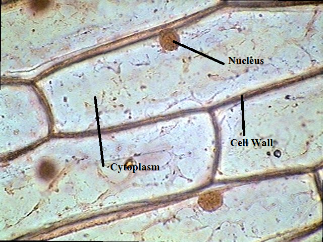
Onion cell cells microscope micrograph under 40x labeled stock alamy microscopic skin magnification root tip allium high epidermis bulb sectionOnion cell under microscope labeled drawing Ncert class 9 science lab manualMicroscope labeled.


Onion Cell Under Microscope Labeled Drawing - apostolicavideo

Onion Epidermal Cell Labeled Diagram - Wiring Diagram Pictures
Biopedia: Practicals

Onion_Cells – BIOBIZNEWS

Onion Cells under Microscope

NCERT Class 9 Science Lab Manual - Slide of Onion Peel and Cheek Cells

draw the figure of an onion peel showing cell - Brainly.in

Onion cell microscope hi-res stock photography and images - Alamy
The Science Scoop: Onion Cell Lab

Figure of onion peel showing cell - Brainly.in